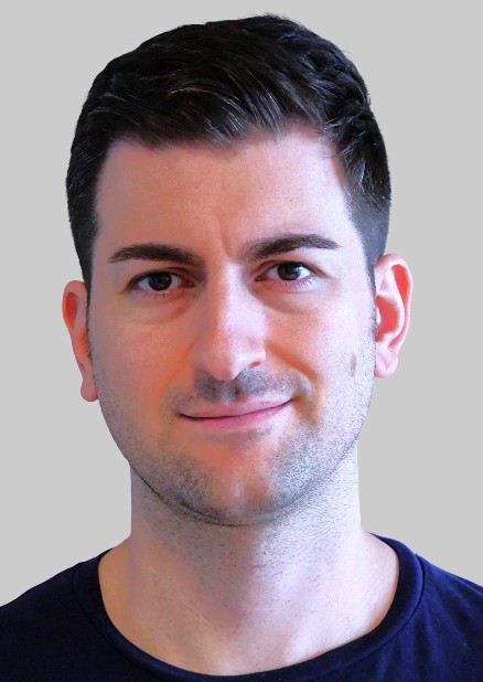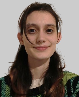

Following my degree in Chemistry, I completed my PhD at the Institut Químic de Sarrià in Barcelona in 2004 under the supervision of Prof. Santi Nonell. During that time, I studied the photophysical properties of phenalenone derivatives, with particular emphasis on singlet oxygen photosensitization, using a range of spectroscopic techniques. In 2005 I moved to the laboratory of Prof. Johan Hofkens at the Katholieke Universiteit Leuven, Belgium, to learn single-molecule and super-resolution fluorescence microscopy. I investigated the photophysical properties of different molecules such as perylene diimide dendrimers and a range of fluorescent proteins.
My most representative result from that period was the single-molecule characterization of the photoswitching properties of the fluorescent protein Dronpa and its mutants. Importantly, we showed how the thorough understanding of photophysics can help optimize super-resolution imaging (Flors et al, J. Am Chem. Soc. 2007). Having gained expertise in a new technique with great potential, I moved to the University of Edinburgh in 2008 to begin my independent research career, funded by the Engineering and Physical Sciences Research Council and The Royal Society. I started a new research program to develop methodology for super-resolution imaging of DNA based on single-molecule localization (Flors et al, ChemPhysChem 2009; Curr. Op. Chem. Biol. 2011). In February 2012 I moved to IMDEA Nanociencia with a Ramón y Cajal fellowship, and I am now Research Professor.
At IMDEA Nanociencia I continue working on the improvement of super-resolution fluorescence microscopy methods, most recently combining them with atomic force microscopy. In parallel to the super-resolution work, I am also interested in the photosensitizing properties of fluorescent proteins and their applications in advanced microscopy and phototherapy.

MEET THE FELLOW
Alvaro Castells Graduated in Biochemistry and Biomedical Sciences (University of Valencia, 2014) and holds a Ph.D. in Biomedicine from Pompeu Fabra University (2019). During his PhD, he worked on the Reprogramming and Regeneration lab in the Centre for Genomic Regulation.
From there, he was hired as an Associate Researcher by the Guangzhou Regenerative Medicine and Health Guangdong Laboratory (GRMH-GDL) (2019-2022) and as an Associate Researcher in the Guangdong Provincial People’s Hospital, Guangzhou, China (2022-2024). Since 2024, he has been working at IMDEA Nanociencia as a MSCA-IDEAL postdoctoral researcher, in the Prof. Cristina Flors lab.
Alvaro´s expertise is centered on the application of Advanced Microscopy Methods to understanding the relation between chromatin structure and cell function. He has a deep understanding of Single Molecule Localization Microscopy and chromatin architecture.
Castells-Garcia, A., Ed-daoui, I., González-Almela, E., Vicario, C., Ottestrom, J., Lakadamyali, M., Neguembor, M.V., and Cosma, Maria P. (2021). Super resolution microscopy reveals how elongating RNA polymerase II and nascent RNA interact with nucleosome clutches. Nucleic acids research.
Otterstrom, J., Castells-Garcia, A., Vicario, C., Gomez-Garcia, P.A., Cosma, M.P., and Lakadamyali, M. (2019). Super-resolution microscopy reveals how histone tail acetylation affects DNA compaction within nucleosomes in vivo. Nucleic acids research 47, 8470-8484.
During my bachelors degree in Chemistry at Complutense University of Madrid, I mainly developed synthetic organic chemistry techniques, having done my bachelor thesis at UCM Pluridisciplinary Institute in the synthesis of fluorescent Ca(II) selective probes. After that, I further developed my skills enrolling in the Interuniversity Organic Chemistry Master, where I finished my master's thesis in the development and application of organic reactions in flow microreactors, at Eli Lilly & Co. There, I performed classic organic chemistry procedures along with ozonolysis reactions using a gas-liquid flow platform, developing new techniques to obtain pharmacologically relevant compounds.
Currently, I am a PhD student cosupervised by Dr Cristina Flors and Dr Nazario Martin at IMDEA Nanociencia. Here I work on the synthesis and advanced characterization of fluorescent glyco-fullerenes with biological properties.

I obtained a degree in Biology from the University of Alicante in 2018 and subsequently completed a Master's degree in Research and Advances in Molecular and Cellular Immunology at the University of Granada, which allowed me to gain experience in proteomic techniques applied to models of chronic inflammation. The challenges imposed by proteomic analysis led me to broaden my knowledge in the development of efficient automation tools by completing a Master's degree in Bioinformatics at the University of Murcia.
Afterwards, I did my PhD in Biomedicine at the Biophysics and Bioengineering Unit of the University of Barcelona under the supervision of Dr. Nuria Gavara Casas. This focused on the use of biophysical biomarkers in combination with machine learning to explore the mechano-pharmacology of fibroblasts in lung cancer on mimetic substrates.
At the same time, I gained experience in atomic force microscopy (AFM) techniques on biomaterials and different biological samples.I am now working in the group of Prof. Cristina Flors at IMDEA Nanoscience exploring nanomechanical aspects of bacteria-material interactions.
